
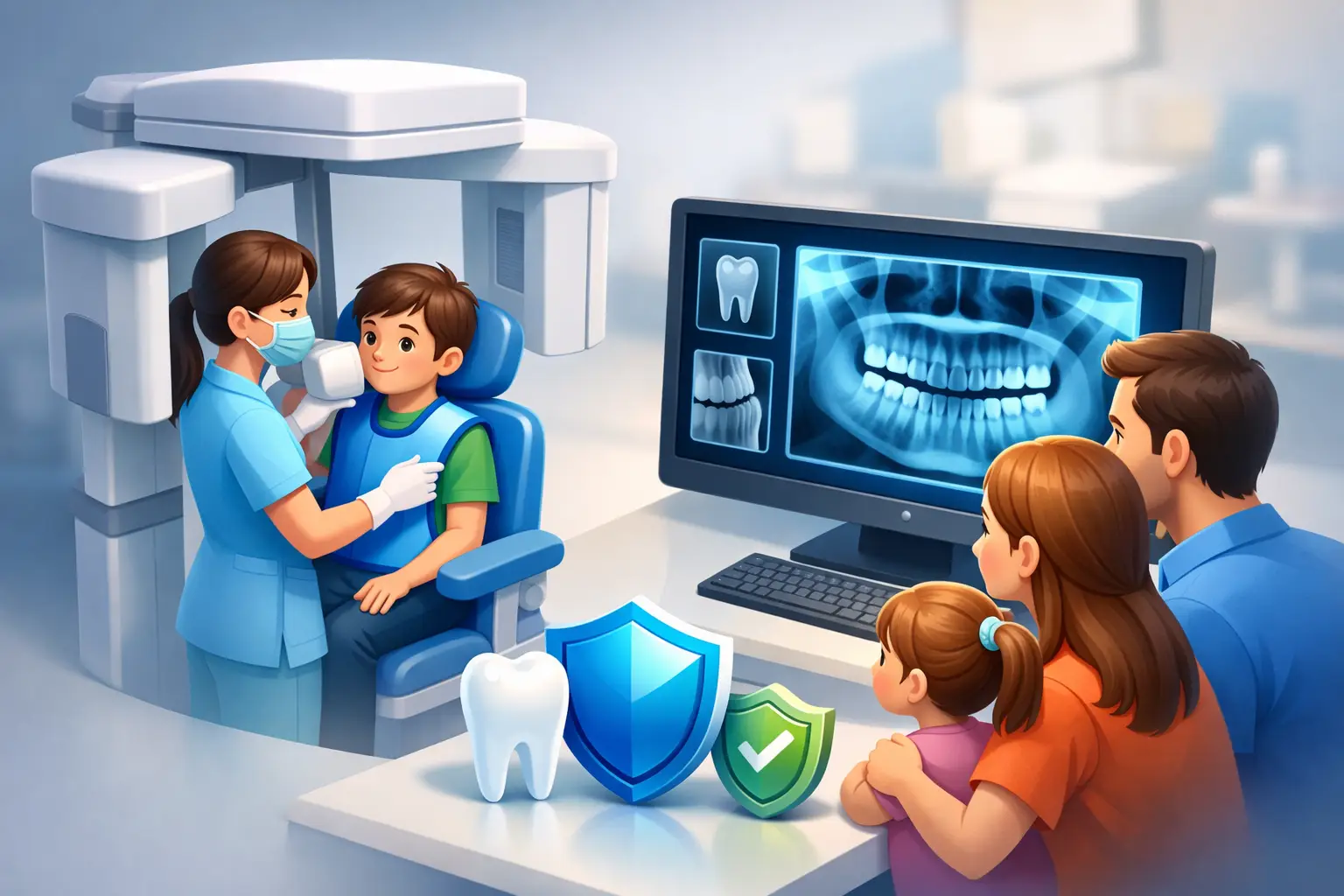

A dental X-ray is not a routine extra added to an appointment. It is a diagnostic tool used when it can answer a question that an examination alone cannot. For families who ask about digital dental x rays safety, the reassuring answer is that modern imaging uses very low radiation doses and is prescribed thoughtfully, based on your individual oral health needs.
At Kingsway Family Dentistry, we believe patients should understand why an X-ray is recommended before it is taken. A clear explanation can make a child’s first dental visit, a parent’s preventive appointment, or an urgent visit for tooth pain feel far more manageable.
Why dentists use dental X-rays
Even a thorough visual examination cannot show everything happening below the gumline or inside a tooth. X-rays help dentists identify concerns early, often before they cause pain or require more involved treatment.
Depending on the type of image, X-rays may reveal decay between teeth, infection near a tooth root, bone changes associated with gum disease, developing teeth in children, impacted wisdom teeth, fractures, and problems beneath existing fillings, crowns, or bridges. They can also help with treatment planning for root canal therapy, implants, orthodontic care, and extractions.
That early information matters. A small cavity found between teeth may be addressed with a conservative filling. The same area, left unseen until it becomes painful, may eventually require more extensive restorative care. The purpose of imaging is not simply to take pictures. It is to support timely, appropriate decisions that protect your comfort and oral health.
Digital dental X-rays safety and radiation exposure
Digital dental X-rays use electronic sensors rather than traditional film. These sensors are highly sensitive, so they generally require less radiation to create a clear image. The image appears quickly on a screen, allowing your dental team to review it with you and adjust contrast or magnification when helpful.
Radiation is part of many aspects of daily life. We are exposed to small amounts naturally from the environment, and the exact dose from a dental X-ray depends on the image being taken, the equipment used, and the settings needed for the patient. A single small intraoral image involves a very low dose. A panoramic X-ray, which captures a broader view of the jaws, has a different exposure level. Three-dimensional CBCT imaging uses more radiation than standard dental images and is reserved for situations where its added detail will meaningfully affect treatment planning.
The key point is not that every image is necessary for every person. It is that modern dental imaging is used selectively. Dental teams follow the principle of keeping radiation exposure as low as reasonably achievable while still obtaining the information needed for accurate care.
How your dental team keeps imaging focused
Safe imaging begins before the sensor is placed. Your dentist reviews your health history, examines your mouth, and considers whether recent images are available and still clinically useful. If you have had X-rays taken at another office, let the team know. In many cases, existing diagnostic images can be reviewed rather than repeated.
X-rays are not scheduled on the same timetable for every patient. Someone with a history of frequent cavities, active gum disease, a new concern, or ongoing restorative treatment may need images more often than a patient with stable oral health and low cavity risk. Children may also need images at particular stages of development, since their teeth and jaws change quickly.
Image type matters as well. A bitewing X-ray is often used to check for decay between back teeth. A periapical image looks closely at an individual tooth and its root. A panoramic image gives a wide view of the teeth, jaws, and surrounding structures. Your dentist should choose the smallest number and type of images that will answer the clinical question.
Modern equipment, properly maintained sensors, careful positioning, and trained staff all contribute to image quality at a low dose. Clear images prevent the need for unnecessary retakes. If an image must be repeated because a child moved or a sensor shifted, the team will explain why and take steps to make the next attempt more comfortable.
What protective measures may be used
Your dental office will use established radiation-safety procedures, including properly calibrated digital equipment and appropriate exposure settings. Protective shielding may also be used according to the type of image, patient needs, and current professional guidance.
You may be familiar with a lead apron or thyroid collar. These protections have long been common in dental care. Recommendations and regulatory requirements can vary by location as technology and scientific guidance evolve, but patient protection remains the priority. If you have questions about what will be used during your appointment, ask before imaging begins. Your dental team should welcome that conversation.
For children, comfort and positioning are especially important. A calm explanation, child-sized equipment when available, and a few moments to practice can reduce movement and help obtain the needed image efficiently. Parents are often surprised by how quickly digital imaging is completed.
Are dental X-rays safe during pregnancy?
If you are pregnant or think you may be pregnant, tell your dental team. This information helps your dentist consider the urgency of the image and choose the most appropriate approach.
Dental problems do not always wait until after pregnancy. Severe tooth pain, swelling, trauma, or suspected infection may require prompt diagnosis and treatment. In those situations, dental X-rays can be clinically appropriate because delaying care may create a greater health concern. When imaging is not urgent, your dentist may recommend waiting. The right choice depends on your symptoms, stage of care, and the information needed to treat you safely.
When an X-ray is worth it
It is understandable to want to avoid any unnecessary exposure. The best question is not simply, “Do I need X-rays?” but “What will this image help us determine?” A good recommendation has a specific purpose.
For example, an X-ray may help identify the source of sudden sensitivity beneath an old crown, confirm whether an infection is present, monitor bone support around teeth, or guide the care of a child whose permanent teeth are beginning to erupt. Without that information, treatment can become guesswork, or a hidden problem may progress unnoticed.
You are always entitled to ask why an image is recommended, what type of image is being taken, whether prior images can be used, and how the result may affect your treatment plan. Clear answers are part of patient-centered care, not an inconvenience.
A practical approach for busy families
Keep your dental team informed about changes in your health, pregnancy status, recent dental treatment, and any X-rays taken elsewhere. Bring up new symptoms such as swelling, persistent sensitivity, a cracked tooth, or pain when biting, since these details may change the kind of image that is most useful.
For routine visits, remember that imaging is only one part of a comprehensive examination. Your dentist also evaluates your teeth, gums, bite, restorations, oral tissues, and concerns you may have about function or appearance. Together, these findings guide a personalized care plan.
Digital dental X-rays are designed to give your dentist a clearer view while keeping exposure carefully limited. When a recommendation is tied to your specific needs and explained in plain language, imaging becomes what it should be: a small, carefully considered step toward preventing pain and protecting your family’s smiles.